RIKEN Center for Computational Science

Menu
Menu
OVERVIEW 計算科学研究機構とは

Computational Structural Biology Research Unit

Developing New Methods and Computational Tools for Integrative Structural Biology

Biomolecules such as proteins and RNA form large complexes that work together to accomplish core biological functions. Dysfunction of those biomolecules may result in severe diseases. To understand such diseases and develop treatments, mechanisms of these protein functions need to be understood. This requires determining their 3-dimensional structures. However, most biomolecules move and change their shapes upon accomplishing their functions, and such dynamic factors should also be considered.

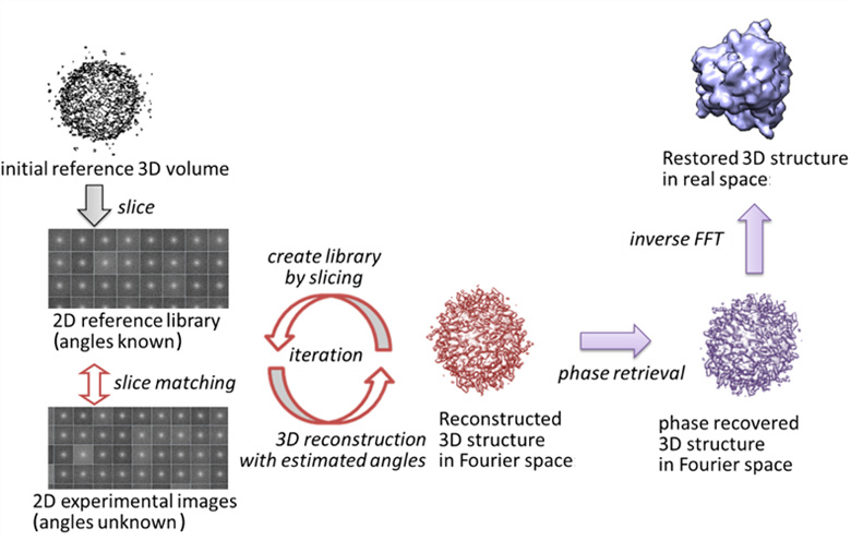
3D structures of biomolecules are obtained primarily through experimental studies. For example, X-ray crystallography provides high-resolution structures. However, it requires crystallization of the biological molecule, which can be quite a challenge. In other methods, such as cryo-electron microscopy (cryo-EM), 2D images of the individual biological molecules are collected, and then be reassembled to form a 3D structure, though generally of a lower resolution. Still, this presents some advantages over X-ray crystallography as large complexes can be studied as well as their dynamics.

Recently, the X-ray free electron laser (XFEL) also opened the possibility to directly image single molecules, using its high intensity X-ray laser. Our interdisciplinary research unit aims to develop computational tools to determine 3D structures and dynamics, utilizing cryo-EM and XFEL experimental data. Such computational tools will be employed on the K and post-K computers to take advantage of the resources for large scale data analyses, and also to share these tools with the scientific community. Applications could provide new insights into the structure and dynamics of important biomolecules that are unattainable with existing techniques.

Recent Achievements

Restoring 3D structures from XFEL diffraction data
Recent development of intense XFEL light sources offers the possibility to obtain new structural information of biological macromolecules, in particular imaging of single macromolecules. Atomic level resolution such as obtained by X-ray crystallography cannot yet be achieved given current experimental conditions. But as XFEL experiments are still undergoing development for routine applications, computational algorithms to understand and analyze experimental data also need to be developed. In particular, to restore the 3D structures of biomolecules from the 2D diffraction patterns obtained by XFEL experiments, computational algorithms are necessary to estimate the laser beam incidence-angles to the molecule.

We are working on a program package for XFEL analysis that is based on XMIPP, commonly used software for image processing of single-particle 3D cryo-electron microscopy. Since XMIPP is designed to work with 2D data in real space, some of the routines were modified to handle 2D data in Fourier space. Through an iterative procedure, the orientations of the biomolecules that each image represents are estimated, and the 3D structure factor-amplitude is reconstructed. The 3D model in real space is obtained by phase retrieval. This approach was successfully tested with experimental data similar to XFEL diffraction patterns taken from a single nanoparticle.

3D structure restoring process

Team Leader Takahito Nakajima

Unit Leader
Florence Tama

Biography: Detail
Annual Report

FY2015 RIKEN AICS Annual Report

(PDF 757KB)
FY2014 RIKEN AICS Annual Report
(PDF 0.98MB)
FY2013 RIKEN AICS Annual Report
(PDF 2.28MB)
FY2012 RIKEN AICS Annual Report
(PDF 436KB)